 | 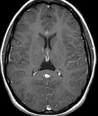 |  |
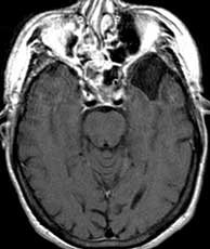
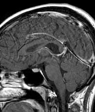
| T1 post-gad | T1 post-gad | SPGR pre-gad |
Diagnosis: Arachnoid cyst of the velum interpositum (presumed)
The velum interpositum is the potential subarachnoid space between the fornix and its attached choroid above and the choroid forming the roof of the 3rd ventricle inferiorly, and is an anterior extension of the quadrigeminal plate cistern just located superior to the pineal gland. If this potential space is simply prominent, it is known as cavum velum interpositum. However, if there is mass effect such as inferior displacement of the internal cerebral veins or the pineal gland, arachnoid cyst is the most likely explanation. Arachnoid cysts are more commonly seen in boys and may present with seizures, headache, or focal neurologic deficit. There is no enhancement in the contents of arachnoid cysts and they follow CSF on all pulse sequences. Over half are located in the middle cranial fossa while up to 10% are located in the suprasellar region and another 10% in the quadrigeminal plate region. The general differential of a non-enhancing CSF containing lesion in this location includes cavum velum interpositum, arachnoid cyst, and epidermoid. Presence of mass effect mitigates strongly against cavum velum interpositum. Likewise, epidermoids tend to engulf surrounding structures rather than produce mass effect making it less likely as well. Related Cases