


Axial CT pre-contrast
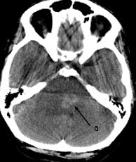

Axial CT pre-contrast Findings: Non enhanced head CT shows a high attenuation lesion involving the subcortex of the left frontal lobe with a large amount of surrounding low attenuation. Another high attenuation lesion (a) is present near the left brachium pontis effacing the 4th ventricle. A third lesion (b) with low attenuation is present involving the right caudate head.