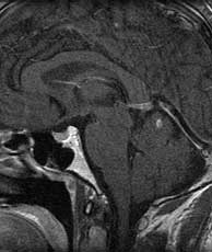
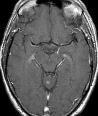
Diagnosis: Small cell lung carcinoma metastases
The brain parenchyma is the most common site of metastatic disease to the CNS. Metastatic disease may also involve the skull, dura, and leptomeninges. The three most common etiologies for brain parenchymal metastatic disease are lung, breast, and melanoma. Brain metastases may represent up to 1/3 of all brain tumors and are multiple in over 80% of patients. The most common location for parenchymal metastatic disease is at the junction of the gray and white matter. Findings of parenchymal metastatic disease to the brain may quite subtle to striking. Metastatic lesions may be solidly or ring enhancing often with a significant amount of surrounding edema and mass effect. Hemorrhage may be seen in metastatic lesions in the brain with any primary; however, renal cell, melanoma, breast cancer, and choriocarcinoma are most likely to produce hemorrhage. The most probable diagnosis in this case, given the imaging findings, is metastatic disease. An atypical presentation of lymphoma is a much less likely possibility, as is cerebritis which could potentially produce multiple small ring enhancing lesions with this distribution early on. This patient was neurological asymptomatic and was imaged as part of his staging workup. Related Cases