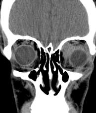

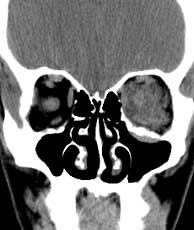
Coronal CT post-contrast
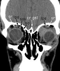
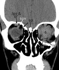
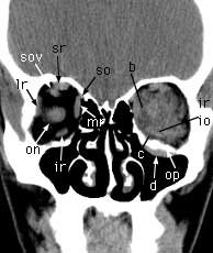
Coronal CT post-contrast Findings: Retrobulbar, intraconal mass (a) with inflammatory changes in the adjacent intraconal fat (b) with extension into the extraconal fat (c). Membrane thickening (d) along the inferior aspect of the orbital process (op) of the maxilla which is not eroded. Extraoccular muscle enlargement is not as apparent as on the axial images. Superior rectus (sr). Levator palpebrae superioris (lps). Superior ophthalmic vein (sov). Superior oblique (so). Crista galli (cr gal). Inferior oblique (io). Inferior rectus (ir). Lateral rectus (lr). Medial rectus (mr). Optic nerve (on).